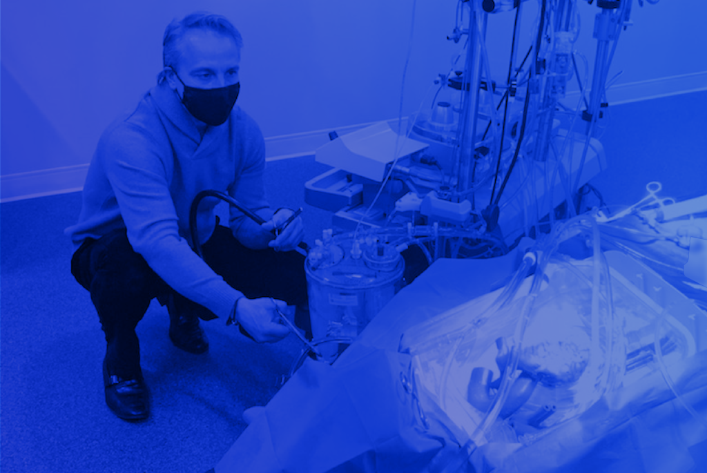
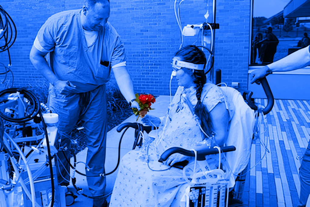
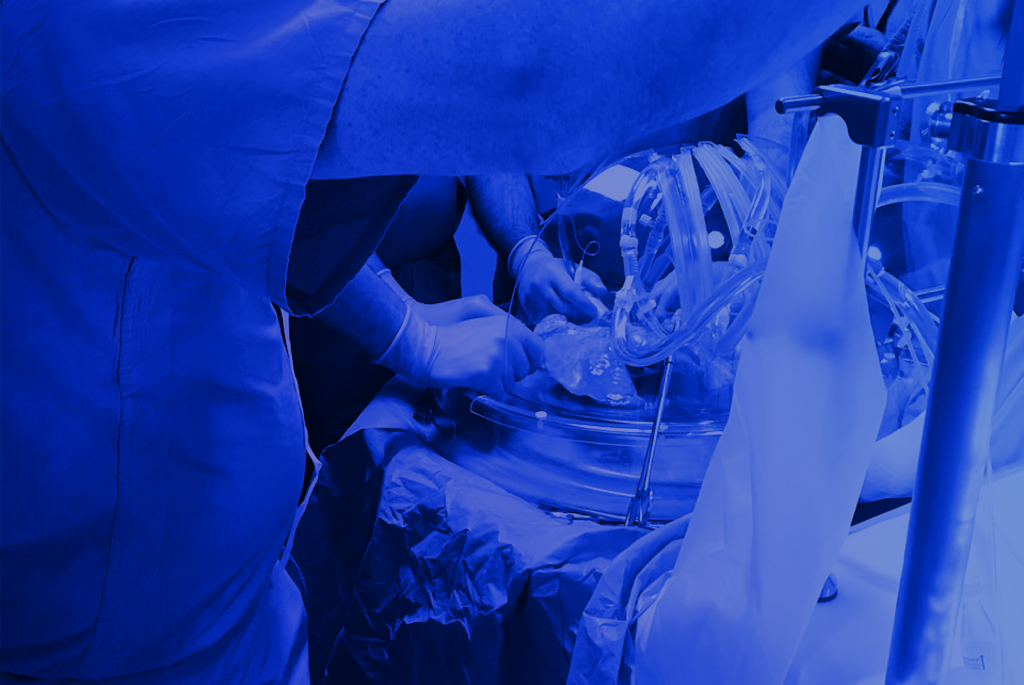
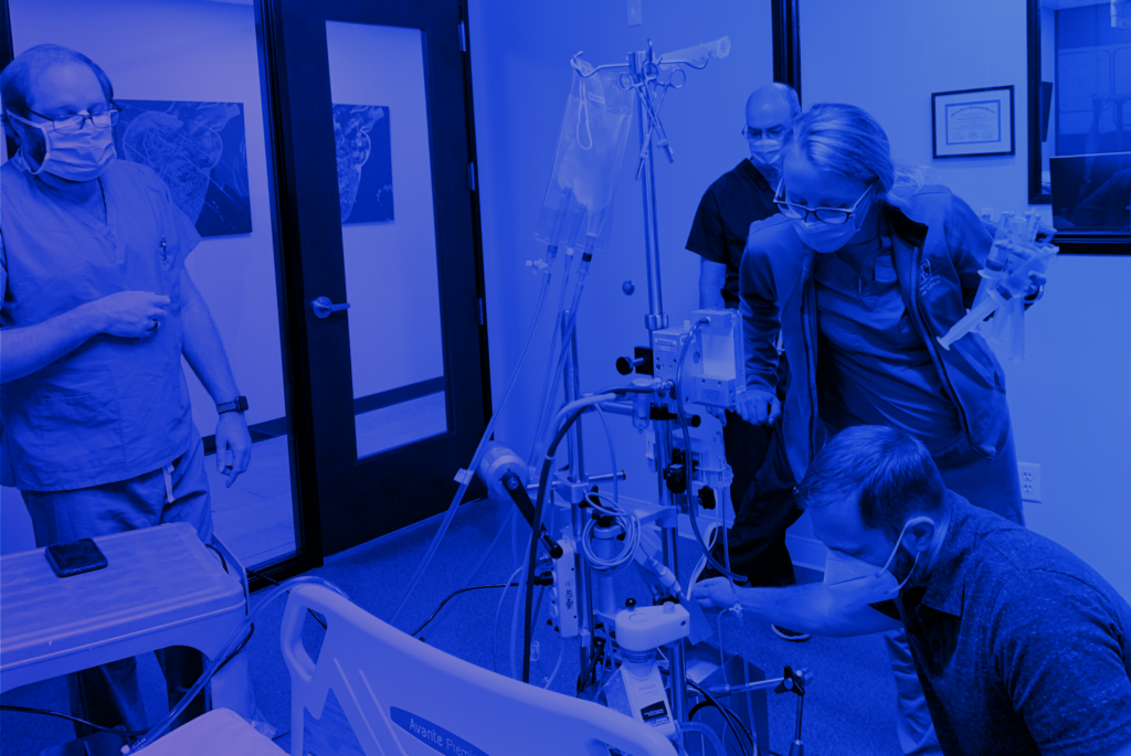
For those with a strong background or interest in healthcare and science, a career working as a perfusionist can be highly rewarding. Perfusionists work as key members of cardiovascular surgical teams by using specialized equipment and patient monitoring techniques. 

Let’s find out more about this exciting career path essential to the practice of modern medicine.

What is a perfusionist?

A perfusionist is a highly skilled, formally trained and accredited healthcare professional that works as a member of an open-heart, surgical team. The perfusionist is responsible for the operation of a cardiopulmonary bypass (CPB) device commonly known as the heart-lung machine. 

During open heart surgery, the patient’s heart is stopped to facilitate delicate surgical techniques. By taking over heart function, the CPB device diverts blood away from the heart and lungs, oxygenates the blood, and then returns the blood to the patient. In addition to operating the heart-lung machine, perfusionists monitor blood circulation parameters and administer blood products or medications to help ensure optimal surgical outcomes. 

In addition to CPB, the perfusionist may apply their skills and expertise to other clinical scenarios, such as:

  • Extracorporeal membrane oxygenation (ECMO) – Similar to CPB used in surgery, this methodology can be used in other scenarios such as acute respiratory failure, cardiac arrest, or as a bridge to heart / lung transplant.
  • Other types of extracorporeal circulation (ECC), such as the delivery of chemotherapeutic drugs to cancer patients’ organs and/or limbs (limb perfusion).

Key perfusionist skills

  • Scientific thought: Perfusion is a demanding medical science that requires skill in understanding and applying scientific data and techniques.
  • Communication: Perfusionists must keep surgeons updated during life saving procedures and critical situations.
  • Stamina: Since surgeries can last several hours, perfusionists require high levels of physical and mental stamina. 
  • Time management: Multiple surgeries, administrative tasks, and patient monitoring all require excellent time management skills.
  • Attention to detail: Perfusionists must pay attention to changes in a patient’s status and make detailed adjustments to care when needed.

What does it take to become a perfusionist?

Most people studying to be a perfusionist already have a solid health and/or science background, such as a Bachelor’s degree in biology or experience as a registered nurse or respiratory therapist. It typically takes two years at an accredited school to complete the requirements for a masters degree in perfusion. 

To begin perfusionist practice, candidates must pass the Perfusion Basic Science Examination (PBSE) and the Clinical Applications in Perfusion Examination (CAPE) to earn their Certified Clinical Perfusionist (CCP) credential. Candidates must complete at least 75 cases, with a minimum of 40 independent cases, before sitting for the exams.

Perfusionists must also satisfy continuing education requirements and maintain up to date knowledge in their field. To maintain credentials, perfusionists complete ongoing training/education and submit proof of education credits to the American Board of Cardiovascular Perfusion (ABCP).

How much does a perfusionist make?

A perfusionist’s work is highly demanding, and one can expect high levels of compensation depending on where you practice, a perfusionist may surpass $95 per hour and starting around $50 per hour.

Conclusion

For those interested in a fast-paced, demanding career in healthcare, perfusion is an attractive option. The standards are rigorous, and the on the job experience can be highly rewarding as you make a difference in life saving patient care.

Article References:

What You Need To Know About Being a Perfusionist

https://www.indeed.com/career-advice/finding-a-job/perfusionist

For hospitals and medical centers, the need for perfusion services continues to increase, and the practice has never been more relevant. Additionally, major shifts in the healthcare industry will also influence practitioners. 

Let’s see how the evolving role of the perfusionist will be impacted by these new trends. 

ECMO Beyond the OR

Traditionally, extracorporeal membrane oxygenation (ECMO), also known as extracorporeal life support (ECLS), was limited to surgical procedures, namely heart bypass. Other procedures, such as heart, lung, and heart/lung transplant have since been added to the situations where qualified perfusionists are indispensable.

Currently, a wide variety of clinical scenarios now involve the use of ECMO/ECLS either as standardized care or as part of clinical investigation, and the list continues to grow. 

Some scenarios where perfusion may be required include:

  • Hypoxemic respiratory failure 
  • Hypercapnic respiratory failure
  • Refractory cardiogenic shock
  • Cardiac arrest
  • Failure to wean from cardiopulmonary bypass after cardiac surgery
  • As a bridge to heart transplantation or lung transplantation
  • Septic shock 
  • Hypothermia
  • Acute respiratory distress syndrome (ARDS)
  • Acute viral pneumonia associated with SARS-CoV-2 infection
  • Transplant organ preservation

Decision Making & Team Skills  

Communication failures are often at the root of adverse events for surgical patients. Increasingly, surgical team integration flattened hierarchies, and standardization is being implemented in an effort to improve outcomes. Checklists, safety briefings, and teamwork training are common techniques used to improve communication in the operating room. 

In the modern OR, the surgeon is no longer the only voice. Rather, surgical teams work as a cohesive unit, and the perfusionist is expected to be a contributing member of the team. In addition to technical expertise, this means developing soft skills, such as clear communication, active listening, and teamwork interaction.  

Impact of Big Data

In healthcare, the rapid expansion of Big Data methodology is part of an effort to enhance research capabilities, reduce costs, and improve outcomes. 

Many hospitals across the country are transitioning their traditional databases to Big Data infrastructure. Advanced data capture and categorization tools such as natural language processing (NLP) and optical character recognition (OCR) are being developed for clinical and research use. Meanwhile, the Internet of Things (IoT), in the form of wearable technology and connected sensors, serves as a key source of data. 

As cardiothoracic surgeons seek ways to innovate, novel approaches to data acquisition and analysis will be part of the new reality. For perfusionists, there is a constant relationship with clinical data that can be collected and analyzed. In the evolving role of the perfusionist, there will be a required understanding of how digitalization impacts clinical and research activity. 

Article references:

AmSECT’s Task Force on the Expansion of Perfusion Scope of Practice

https://www.amsect.org/page/scope-of-practice

30 to 39 pct of severe COVID-19 patients discharged from Wuhan hospitals: official

http://www.xinhuanet.com/english/2020-02/15/c_138785933.htm

Interprofessional communication in the operating room: a narrative review to advance research and practice

https://link.springer.com/article/10.1007/s12630-019-01413-9

Advanced-Data Analytics for Clinical Research Part I: What are the Tools?

https://journals.sagepub.com/doi/abs/10.1177/1556984520902783?journalCode=inva

Category

Perfusion Problems: Complications Associated with ECMO/ECLS

Extracorporeal membrane oxygenation or ECMO (also known as Extracorporeal Life Support or ECLS) supports patients during life-threatening situations, such as heart or lung failure due to illness, after cardiac surgery, or as a bridge for patients awaiting a lung transplant or heart assist device. 

Despite ECLS being used for decades, perfusion problems continue to exist and are associated with significant morbidity and mortality. 

Complications of ECMO

Some of the most serious perfusion problems include vascular, neurological, renal, hemorrhagic complications, and infection. Vascular complications (both bleeding and thrombosis) remain the leading causes of morbidity and mortality in patients treated with ECMO. Survival rates for patients following ECMO therapy in the treatment of cardiogenic shock and cardiac arrest have been reported between 20% and 65%.

Limb Ischemia

During peripheral veno-arterial ECMO, large bore femoral arterial cannulation and hemodynamic instability expose limbs to significant risk for thromboembolic complications. Limb ischemia risk factors include: 

  • Larger cannulas (>20 Fr)
  • Women and younger patients
  • Presence of peripheral arterial disease

Ways to mitigate ischemic risk include vascular evaluation with Doppler ultrasound and near infra-red spectroscopy (NIRS). Some experts recommend a baseline NIRS (SpO2) of greater than 40 and a distal perfusion pressure of 50 mmHg to prevent limb ischemia. Distal perfusion catheter (DPC) placement has been shown to improve limb perfusion and reduce the incidence of limb ischemia.

Vascular Injury

Vascular injury is another serious perfusion problem that can lead to dissection, pseudoaneurysm, and retroperitoneal bleeding. These complications typically occur during the placement or removal of arterial cannulas. Vascular complications occur in 7–14% of patients and are compounded by coagulative abnormalities.

Narrow pseudoaneurysms can be managed with ultrasound-guided thrombin injection. Larger pseudoaneurysms may require surgical intervention. Meanwhile, arterial dissections are frequently asymptomatic. Dissection with arterial occlusion requires prompt differentiation from thromboembolic occlusion.

Infection

Infection rates at groin cannulation sites range from 7% to 20%. Malnourishment and obesity increase the risk of local infection. Strict aseptic technique during cannula placement, handling, and removal are critical to prevent infection.

Additional Perfusion Problems

Other problems associated with ECMO therapy include:

  • Protamine reaction
  • Hypoperfusion
  • Oxygenator failure
  • Blood clotting within the extracorporeal circuit
  • Line separation
  • Gross contamination
  • Transfusion errors
  • Drug errors
  • Gas embolism
  • Electrical or other equipment failures 

It’s imperative that clinicians develop awareness of these perfusion problems and seek to establish preventive and treatment protocols. 

The Oakland University Perfusion Technology Program and the University of Colorado Health Sciences Center published guidelines (found here) about perfusion problems. This resource could be used as a baseline for perfusion programs to build upon and modify to fit their clinical environment.   

Article references:

Management of vascular complications of extra-corporeal membrane oxygenation

https://www.ncbi.nlm.nih.gov/pmc/articles/PMC6039794/#:~:text=Major%20complications%20of%20ECMO%20include,of%20mortality%20in%20ECMO%20patients

Complications of extracorporeal membrane oxygenation for treatment of cardiogenic shock and cardiac arrest: a meta-analysis of 1,866 adult patients

https://pubmed.ncbi.nlm.nih.gov/24210621/

Ten Common Perfusion Problems: Prevention and Treatment Protocols https://amsect.smithbucklin.com/JECT/PDFs/1987_volume19/issue3/ject_1987_v19_n3_schabel.pdf

Category

Mechanical Perfusion and Oxygenation for Extended Ex Vivo Organ Preservation

Since the early days of transplant medicine, the parallel practice of organ preservation branched off into a separate science and field of research. The primary goal of mechanical perfusion and oxygenation is to protect organs from the consequences of hypoxia for as long as possible until transplantation can occur.

Cooling is an obvious method to reduce metabolic demand. The ability to apply appropriate cold temperature ex vivo, in ways that could be readily reversed, has led to considerable research and development. Now, flush cooling and ice storage with synthetic preservation solutions have become the most widely used method and have allowed for cost-effective, multiple retrievals from a single donor. 

For years, static cold storage has dominated clinical preservation practices, but currently, there is growing interest in more dynamic strategies that implement perfusion. Although this method is by no means new, current interest is driven by changing donor demographics, new potential clinical applications, and the need to utilize all available organs for transplantation to meet rising demand.

Perfusion Based Organ Preservation Research

Organ preservation, using continuous mechanical perfusion with blood-based or bloodless perfusates has been an active field of research for years. Beyond simply extending its viability beyond 24 hours (the standard for cold-storage techniques), the research reveals other benefits that perfusion methods might offer.

One recent study out of the Department of Surgery and Transplantation, Swiss Hepato-Pancreato-Biliary (HPB) Center, University Hospital Zurich, Zurich, Switzerland reveals that integrated mechanical perfusion and oxygenation can preserve injured human livers for one week.

Study Design 

Given that organs can usually be delivered within 24 hours to the operating table, what are the advantages of preserving them for longer time frames? The authors of the Zurich study highlight there is a high reject rate due to damage or injury. However, if the body parts could be kept viable and nursed to recovery, they could potentially increase the pool of donors.

The study included the development of specialized perfusion and oxygenation equipment equipped with intravascular pressure and blood gas tension monitoring in addition to a dialysis unit for physiologic electrolyte balance and removal of metabolic waste products from the blood. 

An algorithm was used to automatically adjust the dialysate flow, controlling the concentration of red blood cells (hematocrit) based on continuous measurements. Automated insulin and glucagon administration was used to maintain physiological blood glucose levels (targeted range of 3.5–6.5 mmol l−1).

To simulate diaphragmatic liver movement in the ex vivo system, they were placed on a silicone mat. An inflatable balloon was positioned beneath the mat and connected to an air oscillator to induce movement that mimics diaphragm motion which in turn helps prevent pressure necrosis.

Mechanical perfusion: Study Results & Potential Clinical Applications

After seven-day perfusion, six of the human livers showed preserved function as indicated by bile production, coagulation factor synthesis, maintained cellular energy (ATP), and an intact structure.

As the authors state, with additional research, multiple potential benefits could be realized with extended organ perfusion technology, such as:

  • Repair of damaged or diseased organs to be transplanted later.
  • Graft immunogenicity modification to induce tolerance. 
  • Treatment of tumors with chemotherapeutic agents that are too toxic for systemic use.
  • Safe transport over long distances to improve global sharing.
  • Liver regeneration, divided into segments to regrow ex vivo. Such an approach could double or even triple availability. 
  • Partial grafts for auto-transplantation in patients with liver cancer, obviating the need for immune suppression.

Article References:

Organ Preservation into the 2020s: The Era of Dynamic Intervention

https://www.karger.com/Article/FullText/499610#ref19

An integrated perfusion machine preserves injured human livers for 1 week

https://www.nature.com/articles/s41587-019-0374-x

Category

Utilizing qualitative research to improve clinical outcomes.

The fundamental purpose of qualitative research is to understand better why a particular group behaves as it does. The numbers produced by quantitative research only scratch the surface of these types of questions, being more about learning which sub-groups are essential and how much they may vary in behaviors and responses. In comparison, qualitative research is much better at getting at the ‘hows’ and ‘whys’ that are so important to in-depth knowledge.

First, you select a particular population to study. It can be any group with some level of shared experience. Second, you need to collect some free-form data from (or about) a representative sample of that population. Third, you want to record any potentially relevant information about the sources of that data: age, ethnicity, sex, previous ailments – you name it. Fourth, it is time to highlight the most relevant sections of that qualitative data, have a preliminary perhaps coding each excerpt for the degree to which it supports or refutes a particular theme. These themes can be questions you now want to investigate upfront or questions that the excerpts themselves suggest during your study. This step will be based mainly on the project’s research methodology. If any CCS perfusionists are interested in formulating a study or wish to discuss examining a specific study group, please reach out to Professor Gunaydin or me at any time.

We thought this week’s topic would cover our recent pediatric study and publication, and we hope you enjoy the reading. A STRUCTURED BLOOD CONSERVATION PROGRAM IN PEDIATRIC CARDIAC SURGERY: More Is Better”

Background: The limitation of alternative transfusion practices in infants increases the benefits of blood conservation. We analyzed the efficacy of a structured program to reduce transfusions and transfusion-associated complications in cardiac surgery

Methods: Our pediatric surgery database was reviewed retrospectively, comparing outcomes from two different time periods, after the implementation of an effective blood conservation program beginning in March 2014.  A total of 214 infants (8.1±3.4 months) who underwent biventricular repair utilizing CPB (Group1-Blood conservation) were studied in 12 months (March 2014-February 2015) after the implementation of the new program and compared with 250 infants (7.91±3.2 months) (Group 2-Control-No blood conservation) of the previous 12-month period (March 2013-February 2014).

Results: The proportion of patients transfused with red blood cells was 75.2% (N=188) in the control group and reduced by 16.4% in the study group (58.8%- 126 patients, p <0.01). The mean number of transfusions was 1.25 ± 0.5 units per patient in the control group and decreased to 0.7 ± 0.5 units per patient after the start of the program (P = 0.035). Cerebral oximetry demonstrated better follow-up during the operative period confirming less hemodilution in Group 1. Respiratory support, inotropic need, and ICU stay were significantly better in the study group.

Conclusion: These findings, in addition to attendant risks and side effects of blood transfusion and the rising cost of safer blood products, justify blood conservation in pediatric cardiac operations. Circuit miniaturization, ultrafiltration, and reduced postoperative bleeding, presumably secondary to higher fibrinogen and other coagulation factor levels, contributed to this outcome.

Kevin McCusker Ph.D., MSc., CCP
Assistant Professor of Surgery
New York Medical College
Valhalla, New York 10575                                                          

Serdar Gunaydin M.D., Ph.D.
Professor of Surgery
University of Health Sciences
Ankara, Turkey